Facilities

University Science Park

Expand All

Micron logo

In Micron Bioimaging Facility we offer a full suite of facilities and services to enable biological researchers to access the latest microscope technologies.

The facility team provide training and support on a wide range of microscopes ranging from confocal and widefield to super-resolution imaging. We are happy to advise users on what system is most appropriate for their experiment and to offer help with sample preparation and experimental design. Micron also provides access to the latest image analysis, data storage and visualisation methods including super-resolution quality control, de-noising and machine learning-based image analysis.

The facility in Biochemistry is partnered with many imaging facilities in other departments across Oxford through the Oxford Bioimaging Network, so users have access to the full range of the microscopes that biologists need, from wide-field deconvolution, confocal, light-sheet and 4Pi to 3DSIM, STED, dSTORM, TIRF, FCS and CLEM.

Dunn school logo

Based in the Sir William Dunn School of Pathology, the Dunn School Bioimaging Facility provides Oxford University students and researchers with state-of-the-art preparation, imaging and analysis instrumentation to facilitate their research.

JR Hospital Research Campus

WIMM logo


The Wolfson Imaging Centre has a wide range of advanced microscopes, offering imaging solutions fitting most research needs. From widefield microscopy to the most advanced STED super-resolution and photon-counting technology (FLIM), all researchers across Oxford can have access to hardware working in optimal condition and the expertise needed to profit from it. The facility team also strives to implement new imaging protocols and techniques in a collaborative manner.

                                                                                                            Wolfson Imaging Centre Webpage

The centre currently has the following instruments for use:

  • Leica SP8 STED FALCON inverted confocal
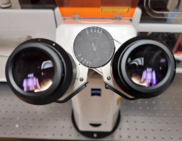
    Look into my eyes
  • Leica Thunder inverted widefield microscope
  • Leica SPE inverted LSM confocal microscope with widefield capabilities
  • Zeiss 780 upright LSM confocal microscope with Insight X3 multi-photon laser
  • Zeiss 880 inverted LSM confocal microscope with Airyscan 1
  • Zeiss 900 upright LSM confocal microscope with Airyscan 2
  • Zeiss 980 inverted LSM confocal microscope with Airyscan 2
  • Zeiss inverted Spinning disk confocal
  • Zeiss Axioscan automatic slide scanner (brightfield and fluorescence widefield)
  • Miltenyi BLAZE Ultramicroscope for large cleared samples
  • DeltaVision Elite inverted widefield system
  • Olympus inverted IX83 TIRF microscope
  • High-end PC workstation with Zeiss ZEN software for Airyscan processing and Image analysis
  • High-end PC workstation with IMARIS 10.0 Image analysis software

Old Road Campus

kennedy logo

Advanced Microscopy in the Kennedy brings together leading expertise and technology, to both use and develop cutting-edge microscopy methods. Made up of the Oxford- Zeiss Center of Excellence in Biomedical Imaging and bespoke homebuilt technologies, the centre champions a culture for respect and innovation in microscopy.

idrm logo
kennedy logo

The DPOC facility is at the forefront of quantifying spatial relationships at subcellular resolutions, complementing the wealth of single-cell RNAseq data and increasing the potential for understanding cellular processes in human disease. Since 2020, the facility has extended to over 70 members testing over 90 biomarkers on 12+ different human tissue types as well as validating 20+ markers on mouse tissues using the Cell DIVE system. Validation of biomarkers for use in 3D imaging will begin with the introduction of the Open-Top Light Sheet Microscope in Q1 2023.

Oxford Brookes University

brookes

The Centre for Bioimaging has a wide range of light and electron microscopes, as well as fully-equipped electron microscopy sample preparation labs. We also have dedicated workstations for 3D image analysis. Our expertise lies in high-resolution live cell imaging and 3D electron microscopy, also known as volumeEM